Using the zebrafish model to develop new treatments for a variety of deadly diseases

Sponsored by

Using the zebrafish model to develop new treatments for a variety of deadly diseases
A different kind of fish story

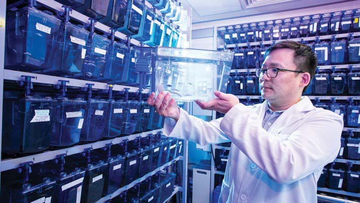
Dr Alvin Ma found zebrafish research of autophagy-related processes in the past could have produced invalid results.
While looking for the causes of leukaemia, Dr Alvin Ma of PolyU made a discovery that could change the way researchers investigate disease
For nearly 50 years, the zebrafish commonly seen in Hong Kong’s tropical fish stores have been prized not so much for their distinctive black and white markings but for their contributions to scientific research. As test subjects, zebrafish are widely used in the search for a better understanding of vertebrate development and human disease.
But now, research findings based on studies of zebrafish embryos have been called into question in a potentially ground-breaking project by a PolyU researcher, Dr Alvin Ma Chun-hang, Assistant Professor in the Department of Health Technology and Informatics.
The role of zebrafish in research

Dr Ma did not originally set out to question the validity of research using zebrafish embryos. Instead, he was focused on investigating the causes of leukaemia, a form of blood cancer. While undertaking this research, he uncovered a problem in the methodology in which zebrafish embryos are employed.
Normally, zebrafish embryos are used as test subjects because they contain blood cell types similar to those found in humans. What’s more, zebrafish are highly fertile, can be kept in large numbers even in cramped laboratories, and are much easier to care for than mice.
What is more surprising, though, is that over 70% of human genes have a zebrafish counterpart, and 82% of human genes related to disease can be found in these tiny striped creatures. Zebrafish embryos are also easy to observe as they are externally fertilised and transparent, which could be maintained using a chemical compound called 1-phenyl 2-thiourea, or PTU.
When observing zebrafish embryos, researchers watch for a process called autophagy, or self-eating. Autophagy is one of the essential processes in living organisms, including humans, as it plays a vital role in ridding cells of unwanted materials when, for example, they are not receiving enough nutrition. It is a vital element in anti-ageing, cell death, tumour suppression and tumour growth.
The problem with PTU
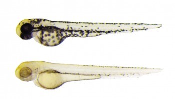
In research using zebrafish embryos, the pigment in the embryos must be supressed in order to increase optical transparency for better imaging of processes such as blood flow. “We treat fish with PTU to inhibit Tyrosinase which is a key enzyme that produces melanin,” Dr Ma said. “By using PTU, the fish embryo will not develop pigment and will be completely transparent.”

Two-day old zebrafish embryos – (top) with pigments in normal development (bottom) transparent after PTU treatment
By making the embryos transparent, however, he discovered a strange phenomenon. “When I tried to suppress the pigmentation in the cells that had been introduced into the embryos, the autophagy levels went up. We tried to troubleshoot what was happening and eventually found that conventional targeting of cells with PTU actually induces autophagy.
“This means that when we are using this model to study any autophagy-related process like cancer, it is a problem.”
The key takeaway from this discovery is that countless studies that have been using PTU in zebrafish embryos in the past could have produced invalid results. “It might mask or interfere with your study,” Dr Ma said.
Setting a new standard
Dr Ma’s discovery was published in the April 2020 issue of Autophagy, the highest impact journal in the field, and has since been frequently cited by peer researchers, receiving a high ranking of 14 out of 193 in the category of Cell Biology.
"The major reason that the journal accepted my paper is that it really tells the field you cannot use PTU anymore — you should avoid using this chemical in autophagy-related research."
As a result of his findings, the chief editor of Autophagy has invited Dr Ma as a co-author in publishing new guidelines on autophagy research using zebrafish embryos, a revision that takes place every three or four years.
But there is still more that needs to be done before we can fully understand how autophagy works. “Autophagy is important for killing cancer cells and plays a key role in developing new treatments,” said Dr Ma. “We now know that autophagy is a much more complex process than we had previously thought.”
With the new guidelines in place, however, Dr Ma is hopeful that other researchers will find better methods of suppressing pigment without affecting autophagy, facilitating the use of the unique zebrafish model and opening the door to new treatments for a variety of deadly diseases, including cancer.