A joint effort: machine learning’s search for the first MRI-based biomarker of osteoarthritis
The University of Queensland has developed a machine learning software tool with Siemens Healthineers that offers a real-world example of AI revolutionising medicine, offering hope for patients with osteoarthritis
Sponsored by

Sponsored by

The knee does not have it easy over the course of a person’s life. As a primary load-bearing joint, it takes our weight and helps us stand, move and keep our balance. When walking, it can take three to four times our body weight, six when running, and that is hard on the protective cartilage in the joint, making it prone to osteoarthritis, for which there is no cure.
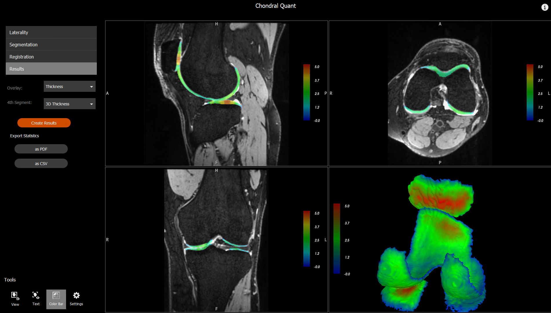
There is much that medical science doesn’t know about osteoarthritis in the knee, but researchers at The University of Queensland (UQ) – in partnership with the CSIRO and Siemens Healthineers – have developed a software tool called Chondral Quant that could change that.
Shekhar Chandra, a doctor in UQ’s School of Electrical Engineering and Computer Science, heads up the research team and says the approach involves two components.
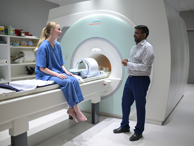
“First, we use the MRI to non-invasively see inside the knee,” he says.
“Then our machine learning algorithm identifies the key aspects of the knee that are important to the disease. We identify the different regions of interest within the knee – the bones, the cartilage and all the bits that are important in osteoarthritis.”

The Chondral Quant software processes the MRI automatically: the algorithm does the heavy lifting and is significantly quicker than its human counterparts.
“Instead of taking hours to do this segmentation of the knee, in our set-up we can turn those hours and hours of analysis per image into minutes – three minutes to be exact,” Chandra says.
This frees up clinicians’ valuable time. It allows them to make better diagnoses and prescribe more suitable treatments, and it also raises hopes that this might lead to the discovery of the world’s first MRI-based biomarker for osteoarthritis.
Chondral Quant’s quantitative analysis not only documents the volume and thickness of cartilage, it allows for a full exploration of its physiology.

It can look at the biochemical makeup of the tissue, assessing how much water is in the cartilage and identifying proteins and other molecules that could be of clinical significance to a condition like osteoarthritis. These can then be explored at scale and speed.
Esther Raithel, senior application developer for MRI at Siemens Healthineers, says the collaboration between Siemens Healthineers, CSIRO, UQ and other university partners enabled the development of the Chondral Quant tool.

“This is a real success story of a partnership between industry and universities, where we could go the whole way from a research topic to a medical product together,” she says.
Feedback data from university partners helped UQ improve the algorithm, while research published in conjunction with UQ’s School of Human Movement and Nutrition Studies supported its regulatory approval.
The Food and Drug Administration has now approved Chondral Quant and the tool can be used in clinical settings in the US, European Union, India, Malaysia, Korea, Switzerland, Brazil, Australia, New Zealand, Turkey and Canada.
“Hopefully we will find that biomarker,” Chandra says.
“Millions of dollars are spent on surgeries, while quality of life is affected due to the pain of osteoarthritis. Hopefully, we can learn enough to prevent that from happening.”
